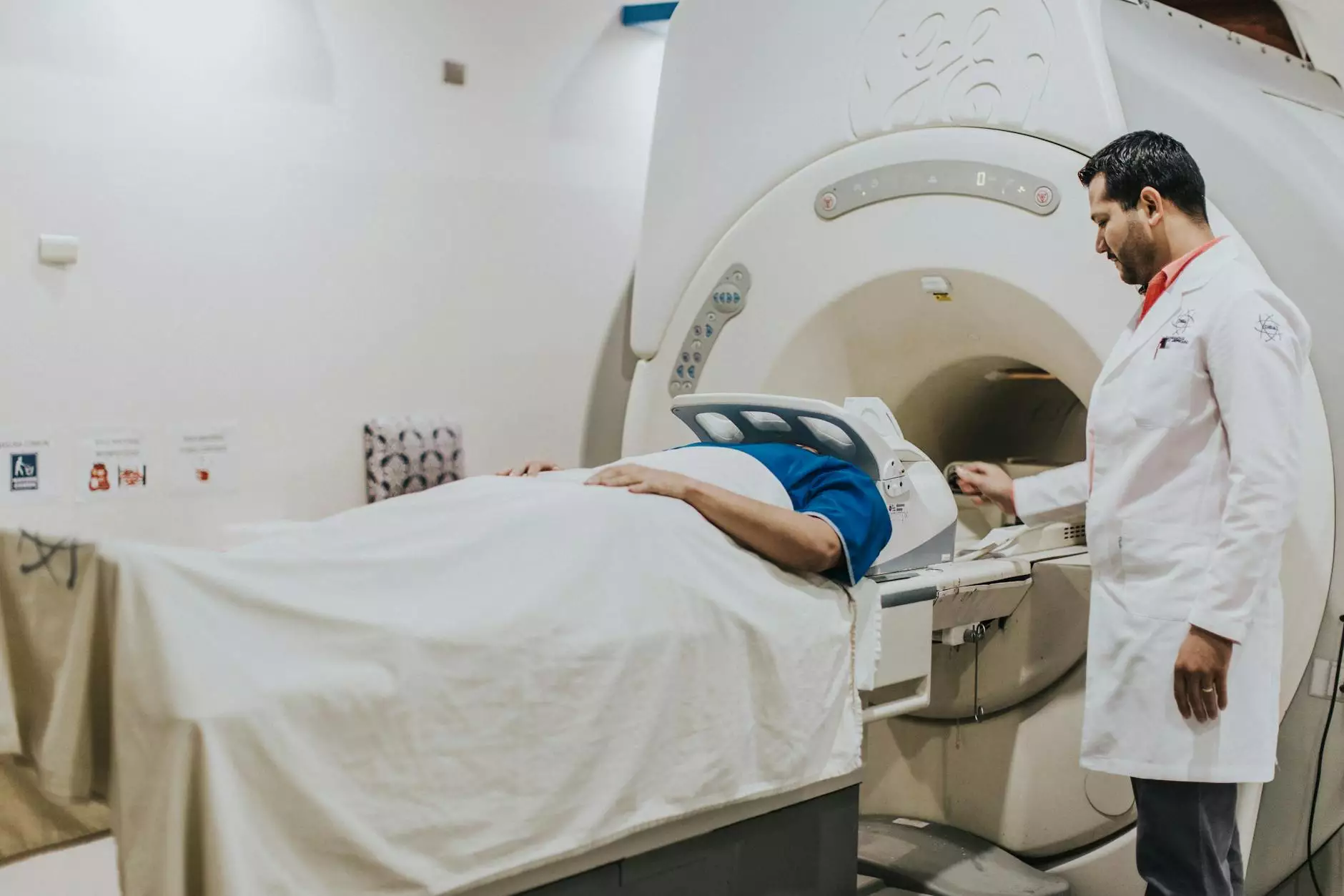
Understanding CT Scans for Lung Cancer Detection

Lung cancer remains one of the leading causes of cancer-related deaths worldwide. Early detection is key to improving survival rates, and one of the most significant advancements in medical imaging is the use of CT scans for lung cancer diagnosis. This article delves into the intricate details of how CT scans function, their benefits, and their role in the broader context of health and medical practices.
What is a CT Scan?
A Computed Tomography (CT) scan is an advanced imaging technique that uses X-rays to create detailed images of the inside of the body. Unlike traditional X-rays, which produce flat images, CT scans generate cross-sectional images (slices) of bones, organs, and tissues. This allows for better visual representation and understanding of the internal structures.
The Role of CT Scans in Lung Cancer Detection
Early Detection
Early detection of lung cancer significantly improves treatment outcomes. CT scans play a crucial role in identifying lung cancers at a stage where they are most treatable. Studies have shown that regular screening with low-dose spiral CT can reduce lung cancer mortality by approximately 20% among high-risk populations, such as smokers and those with a family history of lung cancer.
High-Resolution Imaging
The detail provided by CT scans is unparalleled. These scans offer high-resolution images that can differentiate between lung nodules that are benign versus malignant. Specialized radiologists analyze the lung CT images to assess the size, shape, and density of lung nodules effectively.
CT Scan Techniques
There are various techniques for performing a CT scan, including:
- Low-Dose CT Scans: Designed for screening purposes, these scans minimize radiation exposure while providing quality images for detecting lung cancer.
- Contrast-Enhanced CT Scans: In some cases, a contrast dye may be injected into the bloodstream to enhance the visibility of blood vessels and improve the differentiation between normal and abnormal structures.
- Helical CT Scans: This technique involves a continuous rotation of the X-ray tube, allowing for rapid acquisition of images, which is particularly beneficial in emergency settings.
Limitations and Considerations
While CT scans are invaluable for lung cancer detection, they are not without limitations. Not all lung nodules detected via CT scans are cancerous; many are benign and may require follow-up imaging or biopsies to confirm diagnosis. Additionally, there is a risk of false positives, which can lead to unnecessary anxiety and additional invasive procedures. Furthermore, although advancements in technology have minimized the radiation exposure risks associated with CT scans, patients still need to consider the cumulative exposure from multiple scans over time.
The Importance of Follow-up Procedures
After a CT scan, healthcare providers often recommend follow-up procedures based on the findings. These may include:
- Additional Imaging: Such as PET scans or MRIs to gain further clarity.
- Biopsy: To analyze tissue samples from lung nodules.
- Regular Monitoring: Follow-up CT scans at scheduled intervals to track changes in lung nodules over time.
Integrating CT Scans into Lung Cancer Screening Programs
As part of a comprehensive lung cancer screening program, CT scans are utilized alongside other diagnostic tools to form a complete picture of a patient’s health. This integrated approach ensures:
- Comprehensive Risk Assessment: Evaluating lifestyle factors and genetic predispositions.
- Customized Screening Plans: Designing individualized screening timelines and follow-ups based on patient risks.
- Multidisciplinary Collaboration: Coordinating among various healthcare professionals to facilitate the best care possible.
Advancements in CT Imaging Technology
The field of medical imaging, particularly CT technology, has witnessed significant advancements over the past decade. Innovations such as:
- Low-Dose CT: New technologies have enabled scans with drastically reduced radiation without compromising image quality.
- Artificial Intelligence: AI is being employed to assist radiologists in interpreting scans more accurately, thereby improving early diagnosis rates.
- 3D Imaging: Advances allowing for 3D reconstructions of lung scans provide better visualization and understanding of complex situations.
Patient Experience during a CT Scan
For patients undergoing a CT scan for lung cancer detection, understanding the process can alleviate anxiety:
- Preparation: Patients may be asked to avoid certain foods or medications before the scan.
- During the Scan: Patients lie on a movable table that slides through the CT scanner, and they will be instructed to hold their breath for specific intervals to minimize motion.
- Post-Scan: Once completed, there are usually no recovery times needed, and patients can resume normal activities unless otherwise instructed.
Conclusion: The Future of CT Scans in Lung Cancer Management
As the landscape of lung cancer diagnosis and treatment continues to evolve, the significance of CT scans for lung cancer cannot be understated. These advanced imaging tools not only enhance the accuracy of diagnostics but also pave the way for earlier interventions, personalized treatment plans, and improved patient outcomes. By continuing to invest in research and technology, the medical community aims to improve the efficacy of lung cancer screening and ultimately reduce mortality rates.
For those seeking more information on CT scans and lung cancer detection, or to explore other health and medical services, feel free to visit Hello Physio.


